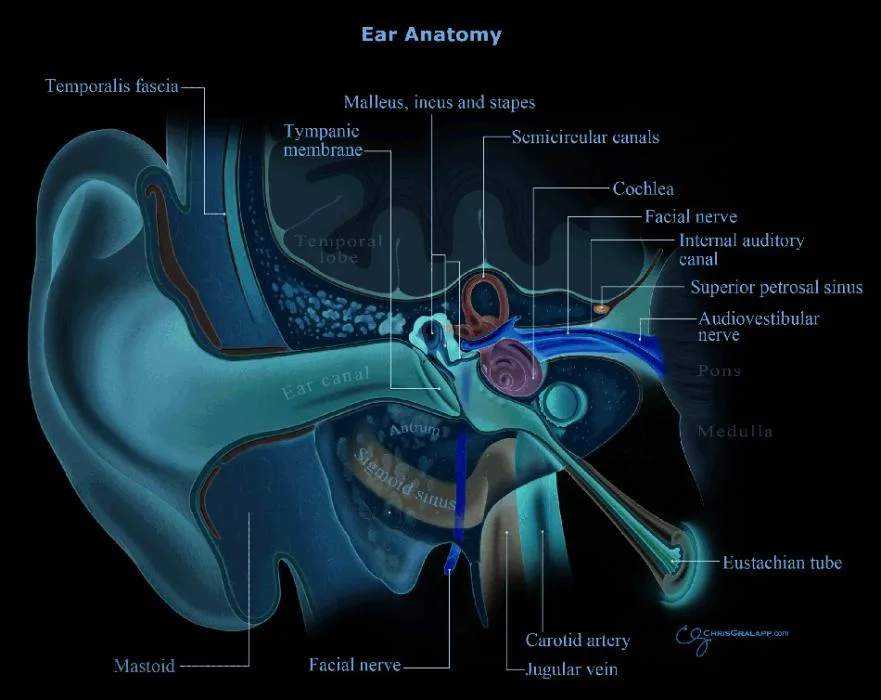
The patulous eustachian tube also known as patent Eustachian tube (PET) is a rare physiological disorder of the eustachian tube, in which a normally closed Eustachian tube stays open intermittently.
PET was first described by Schwartze 1864 when he noted the movement of an atrophic tympanic membrane in synchronous with respiration. Jago described it fully in 1867.
Globally it affects 3 – 6.6% of the general population and is more common in Native Americans, patients with down’s syndrome and preexisting middle ear diseases. The problem affects most commonly females in their third decade of life.
Pathophysiology
Normal eustachian tube has a convexity in the anterolateral wall within the valve area, which flattens during the final stage of the tubal dilation process. In case of PET, there is persistent concavity (defect) in this region of the eustachian tube valve, making it functionally incompatible.
This defect can be due to a deficiency in the lateral cartilaginous lamina, due to thin mucosa or submucosa, reduced ostmann’s fat pad and thin tensor veli palatini muscle, etc.
Etiology
Exact etiology of this condition is still unknown and one-third cases it is considered as idiopathic.
Many conditions are identified as possible associated and causative factors. They include –
- allergic disease – seen in almost 50% of PET patients, chronic inflammation causes patches of mucosal burnout with mucosal and submucosal atrophy in nose, sinuses and eustachian tube.
- rapid weight loss – As little as a six-pound reduction in weight may be sufficient to produce significant tissue atrophy and patulous Eustachian tubes.
- laryngopharyngeal reflux – induce mucosal atrophy
- neurological conditions like multiple sclerosis
- hormonal factors – use of oral contraceptive pills, high estrogen levels in pregnancy especially during the third trimester, anti-androgen therapy for prostate cancer in men
- temporomandibular joint syndrome
- dehydration associated with exercise, caffeine, or diuretics.
- post-surgery – adenoidectomy, cleft palate repair
- Others – Nasal decongestants or cocaine, craniofacial abnormalities, palatal myoclonus, chronic gum chewing.
Pregnancy causes a change in surface tension due to estrogens acting on prostaglandin E, affecting surfactant production.
Clinical features
History
The condition is usually associated with pregnancy, sudden weight loss, rheumatologic disorders or similar chronic illness. There may be previous history of Eustachian tube dysfunction, radiotherapy, multiple Valsalva, laryngopharyngeal reflux, allergy, etc.
Symptoms
These patients usually present with persistent ear blockage, plugged sensation.
They will also have autophony of voice (the patient hears their own breath sounds, synchronous with nasal respiration). The patient complains about hearing his/her own voice and breathing sounds greatly amplified, as if “talking into a barrel.”
Vertigo and giddiness can occur in some patients because of the excessive pressure changes occurring inside the middle ear which gets transmitted to the inner ear via ossicles.
Tinnitus and ear fullness may also be symptoms of PET.
The patient feels relief on supine position or during an upper respiratory tract infection or when doing a Valsalva maneuver. Exercises will worsen the symptoms because of increased respiration.
Signs
Patients with suspected ETD should be examined in a sitting position only. This is because when the patient lies down because mural and intramural pressure in the Eustachian tube increased by venous engorgement making the patient symptomatically better.
On otoscopic examination, in most cases, the tympanic membrane (eardrum) will be atrophic. There will be lateral and medial excursions of the tympanic membrane as patient breaths through the nose. This will get enhanced when the patient breathes with the opposite nostril occluded.
Differential diagnosis
The most important differential diagnosis of the patulous eustachian tube is superior semicircular canal dehiscence syndrome (SCDS). In this situation, autophony will be there, but with no excursions of the tympanic membrane.
Diagnosis
The diagnosis can be done by obtaining a tympanogram while the patient is breathing normally, and a second one while the patient holds his or her breath. Fluctuation in the tympanometric line should coincide with breathing.
The fluctuation can be exaggerated by asking the patient to occlude one nostril and close the mouth during forced inspiration and expiration or by performing a Toynbee or Valsalva maneuver.
In 2018, Kobayashi et al from Japan Otological Society proposed diagnostic criteria for the patulous eustachian tube. This criterias helps in differentiating a definite PET from possible PET.
- Subjective symptoms: One or more of the following symptoms included – autophony, aural fullness, and breathing autophony.
- Tubal obstruction procedures (A or B) clearly improves symptoms
A) Posture change to the supine/lordotic posture.
B) Pharyngeal orifice obstruction procedure (swab, gel, etc) - There are at least one of the following objective findings of PET
A) respiratory fluctuation of the tympanic membrane
B) Variations of external auditory canal pressure synchronized with nasopharyngeal pressure.
C) Sonotubometry shows (1) the probe tone sound pressure level less than 100 dB or (2) an open plateau pattern.
Presence of 1, 2 and 3 constitutes a definite diagnosis of PET, while 1 + (2 or 3) is possible PET. But more studies and validation are needed on diagnostic criteria.
Treatment for patulous eustachian tube
The treatment options for patulous eustachian tubes can be generally classified into three groups – general, medical and surgical management.
General
The general treatment options for patent eustachian tube includes.
- Reassurance – Especially useful in children and teenagers. In this age group patients, this condition is usually self-limited and is probably due to age-related changes, in the structure and function of the Eustachian tube and the adjacent areas, secondary to rapid growth and development.
- If the symptoms are of relatively short duration, the condition may subside without any active treatment.
- Patients can be advised to have weight gain, to avoid diuretics and to adapt a recline or lower head position when symptoms occur, as this will cause congestion of eustachian tube.
Medical management
The medical management of patulous eustachian tubes includes.
- Advice the patient to have good hydration.
- Treatment of any underlying condition
- Nasal irrigation with saline or saline drops.
- Temporary improvements with estrogen nasal topical drops, like Premarin, estradiol is reported – but not FDA approved.
- SSKI (Saturated solutions of Potassium Iodide, 2%), 8 to 10 drops in a glass of juice, taken orally three times daily is found to be useful. This will cause thickening of the mucus.
- Nasal irritant drops containing diluted hydrochloric acid, chlorobutanol, benzyl alcohol, etc. are under trial.
- Avoid the use of nasal steroid sprays, decongestants, and antihistamines which may exacerbate the condition.
Surgery for patulous eustachian tube
Various surgical options are available from different authors with varying success rates. The most commonly practiced ones are –
- Ventilation tubes may be helpful in avoiding membrane excursions with breathing, but not helpful in autophony.
- Teflon injection to para tubal area.
- Transposition of tensor veli palatini muscle by a palatal incision.
- Obstruction of the eustachian tube by a transnasal or middle ear approach is effective, but this may create a late eustachian tube dysfunction necessitating ventilation tube insertion.
- Functional reconstruction of convexity of the anterolateral wall with intraluminal submucosal cartilage implants is under trial and long term results are pending.
- Bluestone practices placement of a plastic catheter into the proximal end of the Eustachian tube – After doing an anterior tympanotomy (opening into middle ear) a small-bore polyethylene tube catheter is inserted into the Eustachian tube orifice, towards the isthmus until it is tightly in place and the flared end is in the middle ear but not touching the malleus. A piece of muscle, fascia, or perichondrium is inserted into the lateral side of the osseous portion of the ET between the catheter and the bony wall of the tube making it secure in position. A tympanostomy tube (grommet / ventilation tube) then inserted into the anteroinferior portion of the tympanic membrane and is kept in place until it is self-extruded.
References
- Oh SJ, Lee IW, Goh EK, Kong SK. Endoscopic autologous cartilage injection for the patulous eustachian tube. Am J Otolaryngol. 2016 Mar-Apr. 37 (2):78-82.
- Bluestone CD, Cantekin EI. “How I do it”–otology and neurotology. A specific issue and its solution. Management of the patulous Eustachian tube. Laryngoscope. 1981 Jan. 91(1):149-52.
- Ikeda R, Kikuchi T, Oshima H, et al. Computed tomography findings of the bony portion of the Eustachian tube with or without patulous Eustachian tube patients. Eur Arch Otorhinolaryngol. 2017 Feb. 274 (2):781-6.
- Kobayashi, T., Morita, M., Yoshioka, S., Mizuta, K., Ohta, S., Kikuchi, T., … Takahashi, H. (2018). Diagnostic criteria for Patulous Eustachian Tube: A proposal by the Japan Otological Society. Auris Nasus Larynx, 45(1), 1–5.
- Doherty JK and Slattery WH: Autologous fat grafting for the refractory patulous eustachian tube. Otolaryngol Head neck Surg 2003;128:88-91
- Ward BK, Ashry Y, Poe DS. Patulous Eustachian tube dysfunction: patient demographics and comorbidities. Otology & Neurotology. 2017 Oct 1;38(9):1362-9.