
There are around 800 lymph nodes in the human body and of these approximately 300 are in the head and neck region. Pathological conditions including tumors, infection and inflammation, etc. of the head and neck region can cause abnormal enlargement of these cervical lymph nodes.
The most common early spread of cancers arising in the head and neck region is to these cervical lymph nodes. Depending on the site of primary cancer (skin, nose and nasal cavity, oral cavity, oropharynx, thyroid, etc.) the sites of lymph nodes involved may vary.
Hence removal of these lymph nodes is an integral part of the surgical treatment of head and neck cancers. The term “neck dissection” refers to en-bloc surgical removal of fibrofatty contents of the neck for the treatment of cervical lymphatic metastases.
Classification of Neck Lymph Nodes
Henri Rouvière was the first to classify cervical lymph nodes in 1932. He classified cervical nodes based upon anatomical landmarks found in dissection, into submental, facial, submandibular, parotid, mastoid, occipital and retropharyngeal nodes, anterior cervical and postero-lateral cervical groups.
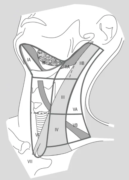
But Rouvière’s system was not suitable for a regular clinical practice where lymph nodes are palpated. Hence came the new classification from Memorial Sloan Kettering Hospital New York in the 1930s, where nodal levels are numbered. This neck lymph node level classification system was modified since then. In 1991, the American Academy of Otolaryngology and Head & Neck Surgery (AAO-HNS) published a standardized version of this classification (Robbins et al) to provide a uniform approach for neck dissection. This was updated in 2002 and 2008 with the addition of sub-levels (IIa, IIb, Va, Vb).
The American Joint Committee on Cancer (AJCC) follows the AAO-HNS classification system for staging of neck nodal metastasis but with an addition of Level VII.
AAO-HNS Lymph node levels
Level Ia: Submental
- Boundaries:
- Superiorly & Laterally: Symphysis menti
- Inferiorly: Hyoid bone
- Medially & Posteriorly: Between the anterior bellies of contralateral and ipsilateral digastric muscles
- Contents: Submental nodes
- Drainage & associated primary malignancies: Drains the skin of the mental region, or chin, the mid-lower lip, the anterior tongue, and the floor of the mouth, mandibular alveolar ridge, and lower lip.
Level Ib: Submandibular
- Boundaries:
- Superiorly: Body of the mandible
- Inferiorly: Hyoid bone
- Anteriorly: Anterior belly of the digastric muscle and mylohyoid
- Posteriorly: Stylohyoid muscle and posterior belly of the digastric muscle
- Contents: Submandibular gland, pre- and post-glandular nodes, and the pre- and post-vascular (fascial artery and vein) nodes, lingual and hypoglossal nerve
- Drainage & associated primary malignancies: Drains other subsites of the oral cavity, anterior nasal cavity, soft tissue structures of the midface, and submandibular gland.
Level II: Upper Jugular
- Boundaries:
- Superiorly: Skull base
- Inferiorly: to the inferior border of the hyoid bone
- Posteriorly: posterior border of the sternocleidomastoid muscle
- Anteriorly: Lateral border of stylohyoid muscle
- Contents: Upper jugular nodes
- Drainage & associated primary malignancies: Posterior oral cavity, Oropharynx, nasal cavity, nasopharynx, larynx, hypopharynx, and parotid.
- Sublevels: Sublevel IIa nodes are located anteroinferior to the vertical plane defined by the spinal accessory nerve. Sublevel IIB nodes are located posterosuperior to the vertical plane defined by the spinal accessory nerve. The clinical significance of this sublevels is that a positive level IIa disease mandates IIb dissection. But elective dissection of laryngeal
and hypopharyngeal malignancy can exclude IIb.
Level III: Middle Jugular
- Boundaries:
- Superiorly: lower border of the hyoid
- Inferiorly: Horizontal plane defined by the inferior border of the cricoid cartilage.
- Anteriorly: Lateral border of the sternohyoid muscle marks the anterior limit
- Posteriorly: Posterior border of the sternocleidomastoid muscle.
- Contents: Middle jugular nodes
- Drainage & associated primary malignancies: Oral cavity, nasopharynx, oropharynx, hypopharynx, and larynx.
Level IV: Lower Jugular
- Boundaries:
- Superiorly: Inferior border of the cricoid cartilage
- Inferiorly: Clavicle
- Anteriorly: Lateral border of the sternohyoid muscle marks the anterior limit
- Posteriorly: Posterior border of the sternocleidomastoid muscle.
- Contents: Lower jugular nodes & Virchow’s nodes
- Drainage & associated primary malignancies: hypopharynx, cervical esophagus, and larynx.
Level V: Posterior triangle
- Boundaries:
- Superiorly: extends from the apex of the convergence of the sternocleidomastoid muscle and trapezius muscle
- Inferiorly: Clavicle
- Anteriorly: posterior border of the sternocleidomastoid muscle
- Posteriorly by the anterior border of the trapezius muscle.
- Contents: Posterior triangle lymph nodes
- Drainage & associated primary malignancies: Nasopharynx, oropharynx, and skin of the posterior scalp and neck.
- Sublevels: Sublevel VA is separated from Sublevel VB by a horizontal plane marking the inferior border of the arch of the cricoid cartilage. Sublevel Va contains a chain of nodes along spinal accessory nerve draining nasopharynx. Sublevel Vb contains nodes related to the thyrocervical trunk which drains the thyroid gland and supraclavicular nodes.
Level VI: Anterior (Central) Compartment Group
- Boundaries:
- Laterally: extends between the two carotid sheaths
- Superiorly: Hyoid bone
- Inferiorly: Suprasternal notch.
- Contents: pre- and paratracheal nodes, the pre-cricoid (Delphian) node, and the perithyroidal nodes, including the lymph nodes along the recurrent laryngeal nerves
- Drainage & associated primary malignancies: thyroid gland, glottic and subglottic larynx, cervical trachea, the apex of the pyriform sinus and cervical esophagus.
PS: The surgical landmark which defines the lateral boundary of Levels II, III, and IV and the corresponding medial boundary of the posterior triangle (Level V) is the plane that parallels the sensory branches of the cervical plexus.
The above lymph node levels classification is not inclusive of several important groups, such as the supraclavicular, parotid, retropharyngeal, and occipital nodes, which is a major limitation of this system.
AJCC Lymph node levels
AJCC lymph node level classification differs from the AAO-HNS by an additional Level of VII. The American Academy considered Level VII to be anatomically mediastinal rather than cervical nodes whereas AJCC consider them along with regional lymph nodes.
Level VII: Superior mediastinal nodes
- Boundaries:
- Superiorly: Suprasternal notch
- Inferiorly: innominate artery
- Contents: Mediastinal nodes
- Drainage & associated primary malignancies: thyroid gland, subglottis, trachea, and cervical esophagus.
References
- Robbins KT, Clayman G, Levine PA, et al. Neck Dissection Classification Update: Revisions Proposed by the American Head and Neck Society and the American Academy of Otolaryngology–Head and Neck Surgery. Arch Otolaryngol Head Neck Surg. 2002;128(7):751–758.
- Robbins KT, Shaha AR, Medina JE, et al. Consensus statement on the classification and terminology of neck dissection. Arch Otolaryngol Head Neck Surg. 2008 May. 134(5):536-8.
- Amin, Mahul B.; Edge, Stephen B.; Greene, Frederick L. AJCC Cancer Staging Manual (Eighth ed.). Springer International Publishing. ISBN 978-3-319-40617-6.