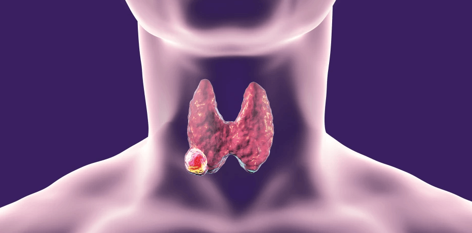
After the diagnosis of any cancer in the body, doctors stage cancer. Staging of thyroid cancers describes how big it is and whether it has spread to any other parts of the body. Staging also helps the doctors in deciding the treatment options, predicting the prognosis and treatment outcomes of cancer.
Most of the cancers are staged using the TNM staging system developed by the American Joint Committee on Cancer (AJCC). Currently, the 8th edition of AJCC staging is followed worldwide with effect from January 1, 2018.
In AJCC staging, TNM stands for primary tumor size and whether it has grown into nearby areas (T), Lymph Nodes affected (L) and spread to other parts of body metastases (M).
Numbers or letters after T, N, and M provide more details about each of these factors. Higher numbers mean more advanced stages of cancer.
Clinical (cTNM) staging is based on inspection, palpation, and imaging (ultrasound, PET / CT, etc.) of the thyroid gland and regional lymph nodes. Pathologic (pTNM) staging is based on all information used for clinical staging along with the surgeon’s description of gross unresected tumor and histopathologic examination report of the resected tumor specimen.
Once T, N, and M categories have been determined, this information is combined in a process called stage grouping to assign an overall stage.
Staging of differentiated thyroid cancer is not like other cancers. This staging system for thyroid cancers takes into account the age of the patient. The breakpoint of age in the AJCC staging system for differentiated thyroid cancer is 55 years of age.
T Staging of Thyroid Cancers
The TNM Staging for differentiated cancers can be summarized as below:
| T Staging for Primary tumor (pT) for Papillary, Follicular, poorly differentiated, Hürthle cell and Anaplastic thyroid carcinomas: | ||
| TX | Primary tumor cannot be assessed. | |
| T0 | No evidence of primary tumor | |
| T1 | Tumor ≤ 2 cm in greatest dimension limited to the thyroid. | |
| T1a | Tumor ≤ 1 cm in greatest dimension limited to the thyroid. | |
| T1b | Tumor > 1 cm but ≤ 2 cm in greatest dimension limited to the thyroid. | |
| T2 | Tumor > 2 cm but ≤ 4 cm in greatest dimension limited to the thyroid. | |
| T3* | Tumor > 4 cm limited to the thyroid or gross extrathyroidal extension invading only strap muscles. | |
| T3a* | Tumor > 4 cm limited to the thyroid. | |
| T3b* | Gross extrathyroidal extension invading only strap muscles (sternohyoid, sternothyroid, thyrohyoid or omohyoid muscles) from a tumor of any size. | |
| T4 | Includes gross extrathyroidal extension into major neck structures. | |
| T4a | Gross extrathyroidal extension invading subcutaneous soft tissues, larynx, trachea, esophagus or recurrent laryngeal nerve from a tumor of any size. | |
| T4b | Gross extrathyroidal extension invading prevertebral fascia or encasing carotid artery or mediastinal vessels from a tumor of any size. | |
| * All categories may be subdivided: (s) solitary tumor and (m) multifocal tumor (the largest tumor determines the classification) | ||
| T Staging for Primary tumor (pT) for Medullary thyroid carcinomas: | ||
| TX – T3 | Definitions are similar to the above | |
| T4 | Advanced disease | |
| T4a | Moderately advanced disease; tumor of any size with gross extrathyroidal extension into the nearby tissues of the neck, including subcutaneous soft tissue, larynx, trachea, esophagus or recurrent laryngeal nerve. | |
| T4b | Very advanced disease; tumor of any size with extension toward the spine or into nearby large blood vessels, invading the prevertebral fascia or encasing the carotid artery or mediastinal vessels. | |
N Staging depends on whether the regional lymph nodes are involved or not,
| N Staging | ||
| NX | Regional lymph nodes cannot be assessed. | |
| N0 | No evidence of locoregional lymph node metastasis | |
| N0a | One or more cytologically or histologically confirmed benign lymph nodes. | |
| N0b | No radiologic or clinical evidence of locoregional lymph node metastasis | |
| N1 | Metastasis to regional nodes | |
| N1a | Metastasis to level VI or VII (pretracheal, paratracheal, or prelaryngeal/Delphian, or upper mediastinal) lymph nodes. This can be a unilateral or bilateral disease. | |
| N1b | Metastasis to unilateral, bilateral, or contralateral lateral neck lymph nodes (levels I, II, III, IV, or V) or retropharyngeal lymph nodes | |
Depending on whether the tumor has spread to other parts of the body like bones, lungs, brain, etc. the M stage is assigned.
| M staging | |
| M0 | No distant metastasis |
| M1 | Distant metastasis |
Once T, N and M stages are calculated the overall prognostic staging is determined. For differentiated thyroid carcinomas other than medullary and anaplastic ones, the prognostic staging is as follows.
| Prognostic staging for papillary, Follicular, poorly differentiated, Hürthle cell carcinoma. | ||||
| When age at diagnosis is… | And T is… | And N is… | And M is… | Then the stage group is… |
| <55 years | Any T | Any N | M0 | I |
| Any T | Any N | M1 | II | |
| ≥55 years | T1 | N0/NX | M0 | I |
| T1 | N1 | M0 | II | |
| T2 | N0/NX | M0 | I | |
| T2 | N1 | M0 | II | |
| T3a/T3b | Any N | M0 | II | |
| T4a | Any N | M0 | III | |
| T4b | Any N | M0 | IVA | |
| Any T | Any N | M1 | IVB | |
For medullary thyroid carcinoma, the staging is as follows.
| Prognostic staging of Medullary Carcinoma Thyroid | |||
| When T is… | And N is… | And M is… | Then the stage group is… |
| T1 | N0 | M0 | I |
| T2 | N0 | M0 | II |
| T3 | N0 | M0 | II |
| T1-3 | N1a | M0 | III |
| T4a | Any N | M0 | IVA |
| T1-3 | N1b | M0 | IVA |
| T4b | Any N | M0 | IVB |
| Any T | Any N | M1 | IVC |
In the case of anaplastic carcinoma, there are only 3 prognostic stages – Stage IVA, IVB, and IVC; basically, means all anaplastic cancers are considered stage IV cancers. Intrathyroidal anaplastic cancers are designated IVA, whereas anaplastic cancers with gross extrathyroidal extension or cervical lymph node metastases are IVB and with distant metastases IVC.
| Prognostic staging for Anaplastic carcinoma | |||
| When T is… | And N is… | And M is… | Then the stage group is… |
| T1-T3a | N0/NX | M0 | IVA |
| T1-T3a | N1 | M0 | IVB |
| T3b | Any N | M0 | IVB |
| T4 | Any N | M0 | IVB |
| Any T | Any N | M1 | IVC |
AJCC 7 vs 8 – Staging of thyroid cancers.
Revision of the system was undertaken to address several specific limitations identified in the 7th edition (AJCC-7), which has been in use since 2009.
The main changes (described in detail below) in AJCC 8th edition can be summarized as
- an increase in the age threshold for defining the high risk of thyroid cancer-related death and
- a decrease in the unfavorable prognostic significance attributed to certain findings (i.e., cervical lymph node metastases and microscopic extrathyroidal extension (ETE), which has been re-defined to include the only invasion of the perithyroidal muscle).
Following are the major changes when comparing AJCC 7 and 8 in the staging of thyroid cancers.
For differentiated thyroid malignancies
- Age cutoff used for staging was increased from 45 to 55 years at diagnosis.
- Minimal extrathyroidal extension detected only on histologic examination was removed from the definition of T3 disease and therefore has no impact on either T category or overall stage.
- N1 disease no longer upstages a patient to stage III; if the patient’s age is < 55 years at diagnosis, N1 disease is stage I; if age is ≥ 55 years, N1 disease is stage II.
- T3a is a new category for tumors > 4 cm confined to the thyroid gland.
- T3b is a new category for tumors of any size demonstrating gross extrathyroidal extension into strap muscles (sternohyoid, sternothyroid, thyrohyoid or omohyoid muscles)
- Level VII lymph nodes, previously classified as lateral neck lymph nodes (N1b), were reclassified as central neck lymph nodes (N1a) to be more anatomically consistent and because level VII presented significant coding difficulties for tumor registrars, clinicians and researchers.
- In differentiated thyroid cancer, the presence of distant metastases in older patients is classified as stage IVB disease rather than stage IVC disease; distant metastasis in anaplastic thyroid cancer continues to be classified as stage IVC disease.
Changes in the staging of Anaplastic carcinoma thyroid are.
- Unlike previous editions where all anaplastic thyroid cancers were classified as T4 disease, anaplastic cancers will now use the same T definitions as differentiated thyroid cancer.
- Intrathyroidal disease is stage IVA, gross extrathyroidal extension or cervical lymph node metastases are stage IVB, and distant metastases are stage IVC.
Compared with the seventh edition, the changes implemented in the eighth edition downstage many patients into lower stages, more accurately reflecting their lower risk of thyroid cancer mortality. The updated system classifies fewer patients as having stage III or IV disease but conveys a poorer prognosis for those who do.
Other systems for staging of thyroid cancers.
Lahey Clinic (Age, Metastases, Extent, Size or AMES Score)
Developed in 1980 with prognostic factors as age, distant metastases, extrathyroidal invasion, and size.
MACIS System
The Metastases, Age, Completeness of Resection, Invasion, Size (MACIS) system was introduced by the MAYO Clinic to eliminate the need for histologic grading of the tumor. The MACIS score is calculated as follows:
- 3.1 (for patients less than 40 years old at diagnosis) or 0.08 x age (if 40 or more years old) plus.
- 0.3 x tumor size (in cm) plus.
- 1 if tumor incompletely resected plus.
- 1 if tumor locally invasive plus.
- 3 if distant metastases present.
In patients with papillary cancer, using the MACIS system, the 20-year, disease-specific mortality for patients with a MACIS score less than 6 was 1 percent; with a MACIS score between 6.0 and 6.99, 11 percent; with a MACIS score between 7.0 and 7.99, 44 percent; and with a MACIS score of 8.0 or more, 76 percent.
Memorial Sloan Kettering (Grade, Age, Metastases, Extent, Size or GAMES)
Published in 1994, this system classifies patients into low-, intermediate-, and high-risk categories.
There are various other, but less popular staging systems also for thyroid malignancies. Names of those staging systems are listed below and more details about each staging system are available from this article.
- European Organization for Research and Treatment of Cancer (EORTC),
- University of Chicago (Clinical Class),
- Ohio State University (OSU),
- Noguchi Thyroid Clinic Staging System (Noguchi),
- University of Münster (Münster),
- National Thyroid Cancer Treatment Cooperative Study (NTCTCS),
- University of Alabama and M.D. Anderson (UAB&MDA),
- Virgen de la Arrixaca University at Murcia (Murcia),
- Cancer Institute Hospital in Tokyo (CIH),
- Ankara Oncology Training and Research Hospital (Ankara)
| Comparison of commonly used risk factors for stratification of risk of death from disease | ||||||||
| Parameters | EORTC
(1979) |
AGES
(1987) |
AMES
(1988) |
MACIS
(1993) |
OSU
(1994) |
GAMES
(1995) |
NTCTCS
(1998) |
TNM
(2017) |
| Patient factors | ||||||||
| Age | Y | Y | Y | Y | – | Y | Y | Y |
| Gender | Y | – | Y | – | – | – | – | – |
| Tumor factors | ||||||||
| Size of primary tumor | – | Y | Y | Y | Y | Y | Y | Y |
| Multicentricity | – | – | – | – | Y | – | Y | |
| Tumor grade | – | Y | – | – | – | Y | – | – |
| Extrathyroidal extension | Y | Y | Y | Y | Y | Y | Y | Y |
| Lymph node involvement | – | – | – | – | Y | Y | Y | Y |
| Distant metastasis | Y | Y | Y | Y | Y | Y | Y | Y |
| Operative factors | ||||||||
| Completeness of resection | – | – | – | Y | – | – | – | – |
| ‘Y’ variable used in defining risk group “-” variable not used. | ||||||||
References
- Casella C, Ministrini S, Galani A, Mastriale F, Cappelli C, Portolani N. The New TNM Staging System for Thyroid Cancer and the Risk of Disease Downstaging. Front Endocrinol (Lausanne). 2018 Sep 18;9:541. doi: 10.3389/fendo.2018.00541.
- Perrier ND, Brierley JD, Tuttle RM. Differentiated and anaplastic thyroid carcinoma: Major changes in the American Joint Committee on Cancer eighth edition cancer staging manual. CA Cancer J Clin. 2018 Jan;68(1):55-63. doi: 10.3322/caac.21439. Epub 2017 Nov 1.
- Tuttle RM, Haugen B, Perrier ND. Updated American Joint Committee on Cancer/Tumor-Node-Metastasis Staging System for Differentiated and Anaplastic Thyroid Cancer (Eighth Edition): What Changed and Why? Thyroid. 2017 Jun;27(6):751-756. doi: 10.1089/thy.2017.0102. Epub 2017 May 19.
- Lang BH, Lo CY, Chan WF, Lam KY, Wan KY. Staging systems for papillary thyroid carcinoma: a review and comparison. Ann Surg. 2007 Mar;245(3):366-78. doi: 10.1097/01.sla.0000250445.92336.2a.