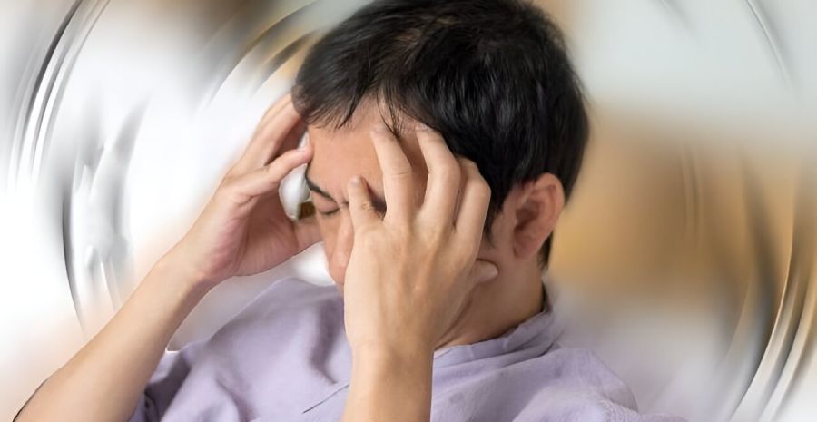
Vestibular neuronitis, also known as acute unilateral peripheral vestibulopathy is a common disorder of unknown origin characterized by inflammation of vestibular nerve (VIII-th cranial nerve) confined with in bony internal acoustic canal, leading to sudden spontaneous isolated total or subtotal loss of afferent vestibular input from one labyrinth.
Vestibular neuronitis is the most common cause of acute onset vertigo of peripheral origin, second to Benign paroxysmal positional vertigo (BPPV).
Causes
- Infections
- Usually viral – neurotropic viruses
- Herpes simplex type I virus can remain dormant in superior and inferior vestibular ganglion. This most commonly affects superior vestibular nerve (SVN). Spontaneous reactivation of the virus can cause neuronitis.
- Following upper respiratory tract infection
- Usually viral – neurotropic viruses
- Vascular
- Frequently spares inferior vestibular nerve, supplied by posterior vestibular artery.
- Involves superior vestibular nerve supplied by anterior vestibular artery.
Clinical features of vestibular neuronitis
- Clinical features are quite typical.
- Single acute onset vertigo, lasting for several days-weeks with gradual resolution.
- A small group of patients presents with similar relapsing episodes which are usually less intense and short lasting.
- Vertigo is so incapacitating in early courses with associated nausea, vomiting and postural imbalance.
- Acute phase last for about 48-72 hours, followed by a period of disequilibrium and sensation of unsteadiness usually lasting for 4-6 weeks.
- Aggravated by head movements; minimized by keeping head still and eyes shut.
- No hearing loss or tinnitus.
Differential diagnosis
- Cerebellar stroke – severe ataxia, signs & symptoms of cerebrovascular accident.
- Negative head impulse test with no catchup saccades in cerebellar stroke.
- Nystagmus is often bi-directional and is not suppressed by visual fixation.
- Patient can’t stand without support even if eyes open; but in vestibular neuritis he can stand.
- Labyrinthine infarction
- caused by occlusion of internal auditory artery, a branch of AICA.
- Entire blood supply to labyrinth is lost causing vestibulopathy with hearing loss. No hearing loss in vestibular neuronitis.
- Autoimmune inner ear disease
- Usually bilateral.
- First attack of Menieres
- Usually may not be associated with hearing loss and tinnitus.
- Patient’s vestibular function recovers soon making it different from vestibular neuronitis.
- Migrainous vertigo
- Acute vertigo associated with multiple sclerosis.
Diagnosis of vestibular neuronitis
- Mainly clinical.
Investigations
- Unilateral vestibular loss
- Spontaneous nystagmus with horizontal and torsional slow components to affected ear and fast component to opposite ear.
- Amplitude increases on looking to normal ear / fast side.
- Head impulse test shows catchup saccades towards affected side.
- Romberg’s / tandem walking / Fukuda or Unterberger – swaying to affected side.
- Subjective visual horizontal test (SVH)
- Single most useful test in vestibular neuronitis.
- The SVH may deviate from true gravitational horizontal by 20 0 or more.
- Electro Nystagmography (ENG) for permanent record of nystagmus.
- Caloric testing not useful in acute setting. Can be used after 3-4 days demonstrating canal paresis.
- MRI Brain
- To rule out central/cerebellar lesions.
- Enhancement of involved vestibular nerve.
- Most commonly SVN is involved – the bony canal through which SVN travels is longer and narrower than that of IVN, making it more susceptible.
- Histopathology of autopsy temporal bones
- Diffuse lymphocytic infiltration with areas of gliosis, reduced number of fibers, marked signs of degeneration in vestibular nerve.
- Loss of sensory hair cells, collapse of ampullary walls are also observed.
Treatment of vestibular neuronitis
- Corticosteroid and antiviral drugs have shown potential benefits.
- Acute phase can be managed by labyrinthine sedatives and other supportive measures.
- Early mobilization and vestibular rehabilitation therapy will improve outcome.
Recovery
- Spontaneous return of vestibular function seen in 50% cases. Time to recovery depends on extend of damage of vestibular nerve.
- Vestibular adaptation – recalibration of motor signals to reduced signals from labyrinth.
- Substitution of other sensory / motor strategies.
- E.g.: rapid eye movement towards lesioned labyrinth.
- Use of visual and proprioceptive information.
- 20% of patients will develop typical posterior semicircular canal BPPV.
Repeated attacks
- In a small proportion of patients, a second attack happens on opposite side – bilateral sequential vestibular neuritis.
- If repeated attacks happen on same side, then Meniere’s disease needs to be considered.