Parapharyngeal abscess is a life-threatening infection, due to spread of infection to the parapharyngeal space.
Epidemiology
- Can occur in any age group
- Most common causes are tonsillitis, peritonsillar abscess, dental infections.
- Suppurative otitis media
- Neck trauma
- Penetrating foreign bodies
- Immunocompromised conditions like HIV, debility etc.
Bacteriology
- Gram negative – Klebsiella pneumoniae > Streptococcus viridans > Pseudomonas aerogenosa (in immunocompromised)
Clinical features
- Similar to peritonsillar abscess – Severely systemically unwell patient with fever, odynophagia (painful swallowing), toxemia, torticollis of neck, trismus (difficulty in mouth opening), airway compromise.
- Decreased oral intake leads to secondary dehydration.
- Maximum swelling in pharynx is more inferiorly placed and behind the tonsil with less edema of palate.
- Tender cervical lymphadenopathy
- The abscess may be felt in the neck which is tender, firm but fluctuant swelling.
- Anterior compartment
- Medially displaced tonsils.
- Trismus due to involvement of pterygoids
- Odynophagia
- Posterior compartment
- IX, X, XI, XII cranial nerve palsies may be present
- Dysphagia, hoarseness, nasal regurgitation, ipsilateral palsies of palate, larynx and tongue.
- Horner’s syndrome due to involvement of cervical sympathetic chain.
Differential diagnosis
- Infections – peritonsillar abscess, Spinal tuberculosis
- Neoplastic – Primary tumors of of parapharyngeal space including deep lobe parotid tumors, carotid body tumors and local spread of tonsil tumors or lymphoma.
- Vascular lesions – Including pseudo aneurysm of internal and common carotid arteries, pseudo aneurysm of lingual arteries (a rare complication in post tonsillectomy).
Investigations
- X-ray neck lateral view – Sometimes a foreign body may be seen.
- Ultrasonogram neck
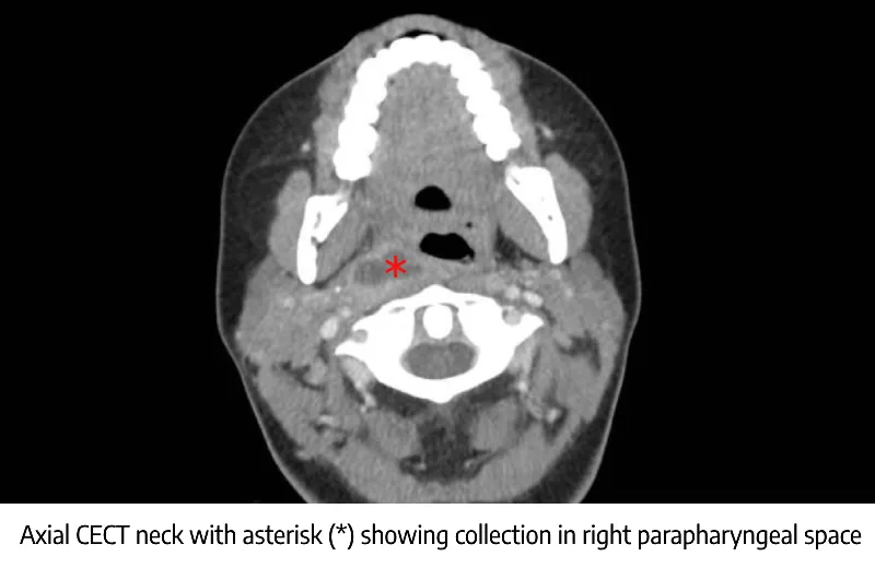
- Contrast enhanced computed tomogram (CECT Scan) of head, neck and chest – Investigation of choice.
- Provides details of the size, location of the abscess and position relative to the large vessels.
- Flexible Nasopharyngo laryngeal endoscopy – to assess the inferior extend of pharyngeal bulge. It also helps to decide whether safe intubation is possible / tracheostomy is needed.
Treatment
- Hospital admission
- Intravenous broad Spectrum antibiotics with anaerobic coverage
- The first-line intravenous antibiotic of choice is amoxicillin with clavulanic acid (150 mg/kg per day) because most of abscesses contain betalactamase producing organisms.
- Anaerobic coverage is provided with intravenous metronidazole (0.5 gm) every 6 hourly.
- Smaller parapharyngeal abscess can be needle aspirated transorally under image guidance through the tonsillar fossa.
- Surgical drainage
- Failure to medical therapy, clinical deterioration etc. are indications for surgical drainage.
- If necessary, a tracheostomy should be done first for securing the airway.
- An awake fiberoptic intubation, is a good option for securing the difficult airway and avoiding a tracheostomy.
- Approaches
- Levitt et al classified surgical approaches into the intraoral and external approaches.
- External drainage can be done by a cervical skin crease incision on side of neck at level of hyoid bone. Depending on the extent of abscess, modified apron incision and hockey stick incision are also considered. Incision is deepened through platysma. Parapharyngeal space is entered by retracting sternocleidomastoid posteriorly by blunt dissection. This step safeguards internal jugular vein and carotid vessels. Abscess cavity is entered and drained.
- Intraoral drainage can be performed under topical anesthesia in the case of accessible abscesses and with patients who are capable of sufficient collaboration. General
anesthesia and orotracheal intubation is needed in small children. Trendelenburg position is adopted to prevent possible aspiration of pus.
- Intravenous broad Spectrum antibiotics with anaerobic coverage
Complications
- Occurs in 16% cases, more common in females.
- Airway obstruction (stridor) due to laryngeal edema or mediastinitis, tracheobronchial aspiration.
- Carotid sheath involvement can result in internal jugular vein thrombosis (Lemierre’s syndrome), internal carotid artery pseudoaneurysm or blow out, Horner’s syndrome.
- Acute pharyngeal perforation, secondary to cervical necrotizing fasciitis.
- Life threatening spread of infection including mediastinitis, retroperitoneal sepsis, airway compromise etc.