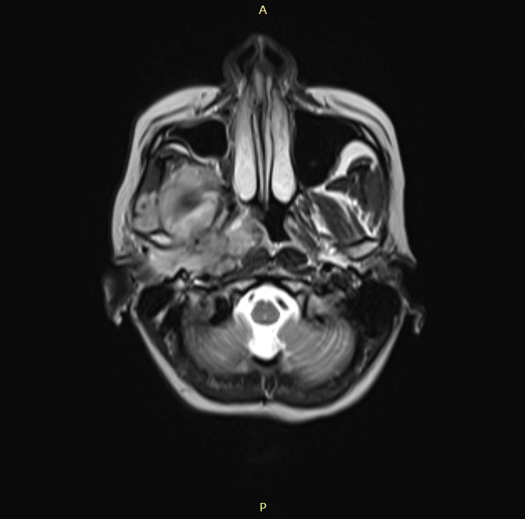
Juvenile nasopharyngeal angiofibroma (JNA) is an uncommon, slow-growing, benign but locally invasive vascular tumor arising from tissues in the sphenopalatine foramen inside the nasal cavity. JNAs are also reported to arise from the posterior aspect of the middle turbinate and rarely from other sites of the nasal cavity and in paranasal sinuses.
JNA tumor was first described by the German surgeon and ophthalmologist Freiherr Maximilian Joseph von Chelius (1794–1846) in 1847 under the name “Fibrous Nasal Polyp”. In 1940 Friedberg introduced the term “angiofibroma”.
Juvenile angiofibroma mostly affects adolescent males of around 14 years of age, also found in children, and some cases reported in the elderly.
Radiological characteristics of JNA are described separately in another article.
Staging systems for juvenile angiofibroma (JA) are important to standardize evaluation, management, and prognosis of the disease. Various surgeons have proposed multiple staging systems but each with their own pros and cons. All of them aimed to eliminate confusion among institutions with regard to surgical approaches, morbidity, and cure rates.
Following are the most popular staging systems for juvenile nasopharyngeal angiofibroma:
Sessions staging
This classification proposed by Sessions et al in 1981 is based on computerized tomographic findings similar to
| Stage IA | Tumor limited to posterior nares and/or nasopharyngeal vault, with no paranasal sinus extension |
| Stage IB | Same as IA but with extension into one or more paranasal sinuses |
| Stage IIA | Minimal lateral extension through the sphenopalatine foramen, into and including a minimal part of the medial-most part of the pterygomaxillary fossa (PMF) |
| Stage IIB | Full occupation of the PMF with anterior displacement of the posterior wall of the maxillary antrum and lateral and/or anterior displacement of the branches of the maxillary artery (superior extension may occur, eroding the orbital bones) |
| Stage IIC | Extension through the PMF into the cheek and temporal fossa |
| Stage III | Intracranial extension |
Advantages
- This was considered to be the first staging system which reflects the unique behavior of JNA more accurately.
Disadvantages
- The classification was based on the number of anatomic sites involved, rather than the actual tumor size which complicates the treatment.
- The classification was based on nasopharyngeal carcinoma staging and thus may result in inaccurate findings.
Fisch staging
In 1983, Fisch proposed the following classification system for JNA
| Type I | Tumor limited to the nasopharynx and nasal cavity, with no bone destruction |
| Type II | Tumor invading the PMF and the maxillary, ethmoid, and sphenoidal sinuses, with bone destruction |
| Type III | Tumor invading the infratemporal fossa, orbit, and parasellar region, remaining lateral to the cavernous sinus |
| Type IV | Massive invasion of the cavernous sinus, optic chiasmal region, or pituitary fossa. |
Chandler staging
Based on the knowledge of presumed site of origin and the growth patterns of JNA, Chandler et al in 1984 proposed the following classification.
| Stage I | Limited to nasopharynx |
| Stage II | Extending to the nasal cavity and/or sphenoid sinus |
| Stage III | Extending into one or more of the following: antrum, ethmoid sinus, pterygomaxillary and infratemporal fossae, orbit, and/or cheek |
| Stage IV | With intracranial extension |
Advantages
- Found to be useful in making decisions about the approach and management of JNA by some surgeons.
Disadvantages
- The complexity of intracranial extension not considered
- Didn’t differentiate extra-nasopharyngeal sites, except the sphenoidal sinus and intracranial cavity.
Andrews-Fisch staging
In 1989, Andrews et al. modified the original Fisch classification, based on the growth pattern of JNA to help surgeons choose access procedures. The modified staging system was named the Andrews–Fisch classification and became the most widely used staging system.
| Class I | Tumor confined to the site of origin at the sphenopalatine foramen may extend unimpeded into the nasopharynx and nasal cavity | |
| Class II | Involvement of the pterygopalatine fossa or the regional paranasal sinuses (the next major sequence of invasion) | |
| Class III | Involvement of the infratemporal fossa or orbital region | |
| Class IIIa | Without intracranial extension | |
| Class IIIb | With extradural intracranial (parasellar) extension | |
| Class IV | Intracranial Intradural extension | |
| Class IVa | Without cavernous sinus, pituitary or optic chaisma infiltration | |
| Class IVb | With the involvement of cavernous sinus/pituitary fossa / or optic chiasma | |
Advantages
- Description of the characteristics of tumor growth and invasion, especially the analysis of extension at the skull base, makes this staging system a practical guide to the surgical approach.
Disadvantages
- As this staging system does not incorporate advances in radiological imaging and surgical techniques, evaluation of the cure rate, the incidence of complications, and local residual and recurrence rates is difficult.
The Fisch system or the Andrews–Fisch system has been recognized as the comprehensive, practical, and applicable guide to surgical approach and prediction of outcome. These systems are the most popular ones used by surgeons and is considered as the most practical and robust one.
Radowski staging
This system proposed by Radkowski et al in 1996 was based on a modification of Session’s system to include the posterior extension to the pterygoid plates and the extent of skull base erosion.
| Stage Ia | Limited to nasal cavity/nasopharynx |
| Stage Ib | Extension into one or more sinuses |
| Stage IIa | Minimal extension into pterygopalatine fossa via pterygomaxillary fissure |
| Stage IIb | Fills pterygomaxillary fossa, bowing the posterior wall of the maxillary antrum anteriorly or extending into the orbit via the inferior orbital fissure without orbital erosion |
| Stage IIc | Infratemporal fossa extension without cheek or pterygoid plate involvement |
| Stage IIIa | Erosion of skull base (middle cranial fossa or pterygoids) with minimal intracranial spread. |
| Stage IIIb | Erosion of skull base with intracranial extension with or without cavernous sinus involvement. |
Advantages
- This staging system takes into account the choice of surgical approach and the risk of recurrence.
Onerci staging
Onerci et al in 2006 proposed this classification system for determining the risk of persistent disease after primary treatment and for choosing the appropriate surgical methods.
| Stages | Features | Residual disease |
| Stage I | Extension to the nose, nasopharyngeal vault, and sphenoidal sinus. | Low possibility of persistent disease by endoscopic/microscopic approach. |
| Stage II | Extension to maxillary sinus or anterior cranial fossa, full occupation of the PMF, limited extension to the infratemporal fossa, or the pterygoid plates posteriorly | Requires more extensive surgery, such as combining Caldwell–Luc or additional endonasal drilling |
| Stage III | Deep extension into the cancellous bone at the base of the body and the greater wing of sphenoid, significant extension to the infratemporal fossa or pterygoid plates posteriorly or orbit region, and obliteration of the cavernous sinus | A high possibility of persistent disease and more extensive surgery |
| Stage IV | Intracranial extension between the pituitary gland and internal carotid artery, extension posterolateral to the internal carotid artery, and extensive intracranial extension | Should be managed via combined extensive (intracranial) surgery |
Limitations
- Cited only in one study.
UPMC staging
With the advancement in endoscopic techniques, along with preoperative image evaluation, and preoperative vascular embolization, tumor size and the extent of sinus disease are less important in predicting complete tumor removal with endonasal surgical techniques. Taking into account of these factors, the University of Pittsburgh Medical Center (UPMC) in 2010 proposed the following classification.
| Stage I | Nasal cavity, pterygopalatine fossa | Can be considered to be minimal tumors and no embolization needed prior to surgery |
| Stage II | Paranasal sinuses, lateral pterygopalatine fossa; no residual vascularity | |
| Stage III | Skull base erosion, orbit, infratemporal fossa involvement; but have no residual vascularity following embolization. | needs prior embolization of the internal maxillary artery and other contributing branches of the external carotid arterial system to devascularize the tumor and facilitate surgery
|
| Stage IV | Skull base erosion, the involvement of orbit, infratemporal fossa; with residual vascularity from the intracranial circulation following embolization | |
| Stage V | With Intracranial extension and residual vascularity following embolization | |
| M – with medial extension (medial cavernous sinus) | ||
| L – with lateral extension (middle fossa) |
Advantages
- Reflects changes in surgical approaches (endoscopic), route of intracranial extension, and extent of vascular supply from the internal carotid artery (ICA).
- more effectively predicts immediate morbidity (including blood loss and the need for multiple operations) and tumor recurrence.
Carillo staging / INCan system
Based on an objective analysis of invasion patterns and tumor size, Carrillo et al proposed a novel and simple classification in 2010.
| Stages | Features | Management |
| Stage I | Tumors located in the nasopharynx, nasal fossae, maxillary antrum, anterior ethmoid cells, and sphenoidal sinus | Can be managed via an endoscopic approach |
| Stage IIa | Tumors invading to pterygomaxillary fossae or infratemporal fossae anterior to pterygoid plates, with major diameter <6 cm | |
| Stage IIb | Tumors invading to pterygomaxillary fossae or infratemporal fossae anterior to pterygoid plates, with major diameter >6 cm | A combined endoscopic and open approach |
| Stage III | Tumors invading to infratemporal fossae posterior to pterygoid plates or posterior ethmoid cells. | |
| Stage IV | Tumors having extensive skull base invasion >2 cm or intracranial invasion | Combined anterolateral skull base approach |
Yi Zixiang staging
Proposed in 2013 by Yi Zixiang et al which divides JNAs into three revised types.
| Types | Features | Approach |
| Type I | Localized in nasal cavity, nasopharynx, sinus, pterygomaxillary fossa. Minimal extension in infratemporal fossa, orbit, or cranial fossa | Transnasal cavity approach with endoscopic guidance |
| Type II | Localized in infratemporal fossa, cheek, deep or minimal anterior cranial fossa extension, minimal middle cranial fossa extension. With or without cavernous sinus and internal carotid artery compression, but dura mater intact | Combined transantral–infratemporal fossa–nasal cavity approach |
| Type III | From pterygomaxillary fossa and superior orbital fissure, extending into middle cranial fossa as a large gourd-shaped lobe | Combined intra- and extracranial approach |
References
- Sessions RB, Nick Bryan R, Naclerio RM, et al. Radiographic staging of Juvenile Angiofibroma. Head Neck Surg. 1981;3:279–83.
- Fisch U. The infratemporal fossa approach for nasopharyngeal tumors. Laryngoscope. 1983;93:36–44.
- Chandler JR, Richard Goulding R, Lee Moskowitz L, Quencer RM. Nasopharyngeal Angiofibromas: staging and management. Ann Otol Rhinol Laryngol. 1984;93(4 Pt 1):322–9.
- Andrews JC, Fisch U, Aeppli U, et al. The surgical management of extensive nasopharyngeal angiofibromas with the infratemporal fossa approach. Laryngoscope. 1989;99:429–37.
- Radkowski D, Mcgill T, Healy GB, et al. Angiofibroma: changes in staging and treatment. Arch Otolaryngol Head Neck Surg. 1996;122:122–9.
- Onerci M, Ogretmenoglu O, Yücel T. Juvenile nasopharyngeal angiofibroma: a revised staging system. Rhinology. 2006;44:39–45.
- Snyderman CH, Pant H, Carrau RL, et al. A new endoscopic staging system for angiofibromas. Arch Otolaryngol Head Neck Surg. 2010;136:588–94.
- Carrillo JF, Maldonado F, Albores O, et al. Juvenile Nasopharyngeal Angiofibroma: clinical factors associated with recurrence, and proposal of a staging system. J Surg Oncol. 2008;98:75–80.
- Yi Z, Fang Z, Lin G, et al. Nasopharyngeal angiofibroma: a concise classification system and appropriate treatment options. Am J Otolaryngol. 2013;34:133–41.