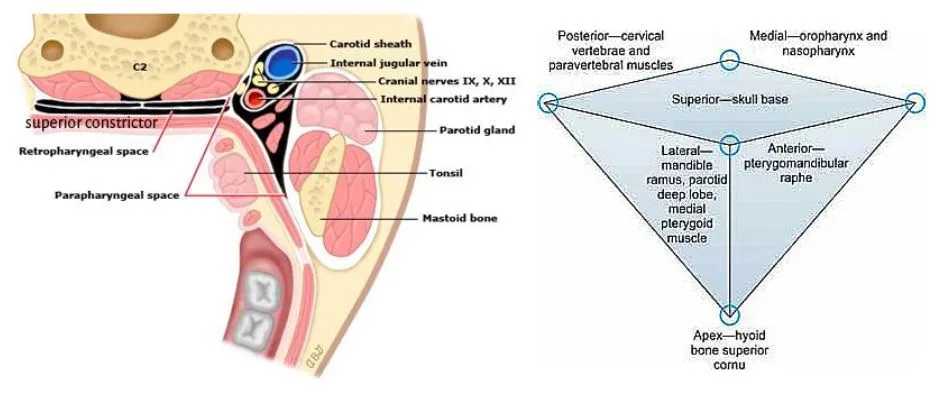
Parapharyngeal space (PPS) is a potential space filled with fat and areolar tissue, lying laterally on either side of pharynx, bounded and subdivided by various condensation of fascia. It is also known as Pterygopharyngeal, Pterygomaxillary, Pharyngomaxillary and Lateral pharyngeal space.
It is an inverted pyramidal shaped space.
- The apex of the space is at hyoid bone below (fascia surrounding submandibular gland).
- The base is at the skull base and tympanic tube above.
- Medial boundary as pharyngo-basillar fascia and superior constrictor muscle.
- Laterally by medial pterygoid muscle, mandibular ramus, parotid gland and parotid fascia.
- Posterior boundary consists of prevertebral structures.

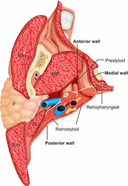
Styloid process with its muscles and condensation of fascia divides the space into a pre-styolid (anterior compartment) and a post-styloid (posterior compartment). More accurately the division is by tensor palati fascial layer.
- Pre-styloid/Anterior compartment
- Apex is at hyoid bone.
- Main contents include pterygoid, tensor palati muscle, fat, deep lobe of parotid.
- Communicates with masticator space.
- Post-styloid
- Continues low into neck.
- Lies posterior and medial to prestyloid compartment.
- Contains carotid sheath (carotid artery, Internal Jugular Vein, Vagus nerve), sympathetic trunk, Cranial nerves IX, X, XII, Major part of Internal Maxillary Artery (IMAX), branches of maxillary nerves and some lymphnodes.
Applied Anatomy of parapharyngeal space
- Pus can collect in the potential parapharygneal space – leading to a life threatening condition known as parapharyngeal abscess.
- Tumors arising in this space involves carotid body tumors, vagal schwannoma etc.
- Removal of parapharyngeal tumors can lead to first bite syndrome.