Sinusitis is one of the commonest causes of patients visit to the oto-rhino-laryngologist (ENT). Making a diagnosis of sinusitis can be challenging as it is not always clear if it is caused by a bacterial infection or by a common cold.
To overcome these limitations, various investigation modalities, like endoscopy, CT scan or MRI) are available for the clinician.
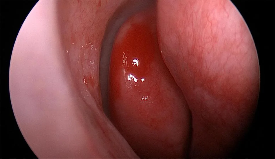
Nasal endoscopy in rhinosinusitis
Nasal endoscopes are tiny telescopes (Hopkin’s rod telescopes) used for looking into the nose to which a special camera can be connected.
Nasal endoscopic examination should be the initial investigation in sinusitis. This allows a detailed examination of the nasal and sinus cavities which is not possible by standard examination such as anterior rhinoscopy using headlight or head mirror.
The doctor can look directly through the endoscope or can use a camera at the end of the scope connected to a larger monitor. It’s a simple procedure which is usually done as an office procedure under local anesthesia.
Advantages of nasal endoscopes
- Most important advantage of nasal endoscopy is that it provides direct and clear field visualization of the nasal cavity and the disease. The angled scopes will even allow visualization of deep structures inside the nasal cavity.
- Nasal endoscopy is a minimally invasive procedure and is completely free of any radiations.
- Nasal endoscopes are easy to handle and the procedure can be done in the office itself which makes it more economical.
- Compared to CT Scan, nasal endoscopy can even detect subtle changes like mucosal hypertrophy, inflammation, erythema, pathologic secretions even before radiographic changes happen. It can differentiate between infective, inflammatory and allergic diseases.
- Endoscopy can be used for other procedures like obtaining cultures and biopsy from the nasal cavity.
Disadvantages of nasal endoscopy
- There are hidden areas inside the nasal cavity that are not well seen by routine nasal endoscopy; most typically the frontal sinuses, spheno-ethmoidal recess, etc.
- Anatomical abnormalities like gross septal deviation etc can make nasal endoscopy difficult.
- Because of the absence of binocular vision, depth perception is not possible with endoscopes.
- There is a good learning curve for mastering the technique of nasal endoscopy.
CT Scan in sinusitis
A computed tomographic (CT) scan of the nose and sinuses can provide very important information in the treatment of patients with chronic sinusitis. At present, CT scan of the nose and paranasal sinuses is considered as the ideal imaging exam (gold standard) to study nasal and paranasal sinuses diseases.
Advantages of CT Scan
- A computed tomographic scan of paranasal sinus is useful in visualizing areas not accessible by routine endoscopy.
- CT Scan can detect mucosal edema and secretions.
- They provide valuable information regarding the complications of sinusitis, its extent and location.
- A preoperative CT scans can act as a road map for the operating surgeon by providing useful information regarding the anatomy of sinuses, osteomeatal complex, and its variations.
Disadvantages of CT
- The main disadvantage with CT scan for the patient is exposed to a large volume of radiations.
- CT Scan represents data at a particular point of time when the disease shows periods of quiescence and exacerbations. This can be followed up by serial endoscopies.
- Though CT scan can detect mucosal edema, thickening, polyps or mass lesions, it fails to differentiate between them and other conditions causing sinus opacification.
- CT Scan provides no idea regarding mucosal hyperemia, mucosal atrophy and other mucosal changes associated with CRS.
- They also provide only minimal information of the type of inflammatory disease like allergic fungal rhinosinusitis, invasive fungal rhinosinusitis, bacterial rhinosinusitis or nasal polyps.
Role of MRI in sinusitis
Magnetic Resonance Imaging (MRI) is another common investigation in sinusitis.
MRI uses powerful magnets and radio waves instead of radiation. These magnets pick up signals from the protons inside the body and are later turned into images. Different types of tissues send back different signals.
T1W MR images can differentiate between the mucosal thickening (will appear isointense) to soft tissue and fluids in the sinuses (appears hypointense). On T2W images, soft tissues and fluids appear hyperintense. Post-contrast, inflamed sinus mucosa shows enhancements while the fluid does not.
Currently, MRI is only used in sinusitis to differentiate soft-tissue structures, such as in cases of suspected fungal infections or neoplasm and when some intracranial complications are suspected.
Advantages
- No radiation exposure.
Disadvantages
- MRI cannot define bony anatomy as well as CT.
- MRI has high false-positive findings, poor bony imaging, and higher cost.
- MRI scans can take a considerably longer time to accomplish than CT scans.
- Difficult to obtain in patients who are claustrophobic.
Best practice
The 2015 American Academy of Otorhinolaryngology and Head & Neck Surgery (AAO-HNS) guidelines strongly recommend that clinicians should confirm a clinical diagnosis of chronic rhinosinusitis (CRS) with objective documentation of sinonasal inflammation.
This can be done via an anterior rhinoscopy, nasal endoscopy, or a computed tomographic scan. When it comes to which method should be used, the following conclusions can be helpful.
Nasal Endoscopy
- In patients who meet guideline symptom criteria for chronic rhinosinusitis, the addition of endoscopic examination can improve diagnostic accuracy for CRS.
- Diagnostic nasal endoscopy should be considered as a routine, early office-based diagnostic tool in the clinical evaluation of CRS and should be performed on all patients suspected of having CRS.
- Nasal endoscopy, a less expensive, easily accessible tool, can reduce the cost of treatment and radiation exposure in a large segment of the population being evaluated for CRS.
- In patients with limited or poor endoscopic visualization, distorted anatomy, due to polyps, or septal deviation or crowding of osteomeatal complex and suspected complications CT scan will be useful in discerning the disease.
CT Scan
- Because of radiation exposure, paranasal sinus CT scans should be obtained judiciously in the appropriate symptom setting.
- CT should not be used routinely to follow the clinical course of CRS because of additive cumulative radiation exposures.
- Sinus imaging could be considered for those patients with refractory symptoms despite maximal medical therapy and in those cases where surgery is being planned.
- The academy recommends against the use of radiographic imaging for patients with acute rhinosinusitis (ARS) unless a complication or alternative diagnosis is suspected.
MRI
- MRI has no advantages over CT scanning in the evaluation of sinusitis.
References
- Bhattacharyya N, Lee LN. Evaluating the diagnosis of chronic rhinosinusitis based on clinical guidelines and endoscopy. Otolaryngol Head Neck Surg. 2010;143:147–151. doi: 10.1016/j.otohns.2010.04.012
- Rosenfeld RM, Piccirillo JF, Chandrasekhar SS, Brook I, Ashok Kumar K, Kramper M, Orlandi RR, Palmer JN, Patel ZM, Peters A, Walsh SA. Clinical practice guideline (update): adult sinusitis. Otolaryngology–Head and Neck Surgery. 2015 Apr;152(2_suppl):S1-39.
- Lohiya SS, Patel SV, Pawde AM, Bokare BD, Sakhare PT. Comparative Study of Diagnostic Nasal Endoscopy and CT Paranasal Sinuses in Diagnosing Chronic Rhinosinusitis. Indian Journal of Otolaryngology and Head & Neck Surgery. 2016 Jun 1;68(2):224-9.