ENT Quiz – Recurrent Meningitis with Hearing loss

Incomplete Cochlear Partition Type I

A 12-year-old male child was referred from the pediatric department following resolution of the third episode of meningitis, with complaints of left-sided hearing loss since childhood.

Parents noticed the hearing loss on the left side when the child was in the fourth standard. No newborn hearing screening was done for the child at birth.

Investigations

  • Blood / CSF / Urine / Immunological studies were normal on presentation to ENT department.
  • Pure tone audiometry – Normal hearing on the right side and profound hearing loss on the left side.
  • Tympanometry – Bilateral Type A curve
  • Distortion product otoacoustic emission – Right present, left absent.
  • Auditory brainstem response (ABR) – Suggestive of profound deafness at left side.
  • MR imaging – No evidence of encephalocele, meningocele, or calvarial defect.
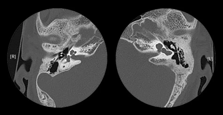
  • High-resolution CT of temporal bones – See the attached image. No evidence of bony dehiscence and the ossicles appeared present and grossly normal.

Question

  • What is your diagnosis from above HRCT temporal bone?


Explanation

The above HRCT temporal bone shows incomplete cochlear partition type I (IP-I) on the left side.

The radiological features are characteristic of incomplete cochlear partition defect – type I (according to the newest classification of cochleovestibular malformations by Sennaroglu and Saatci) or cystic cochleovestibular malformation.

Main CT findings in incomplete cochlear partition type I is

  • a complete absence of entire modiolus
  • absent interscalar septum
  • absent cribriform area resulting in a cystic appearance of the cochlea
  • a large cystic vestibule (sometimes referred as a figure of 8 appearances)
  • wide (most common) or normal cochlear nerve canal.

You can read more about Incomplete partition and radiological characteristics from here.

Recurrent meningitis in an otherwise healthy child should raise suspicion for CSF fistula. Identification of the fistula with prompt control of the leak is necessary to prevent additional episodes of meningitis.

The dictum is that all patients with IP-I and recurrent meningitis with normal tympanic membranes but fluid filling the middle ear and mastoid should have an exploration of the middle ear with special attention to the stapes footplate.

References

  1. Cochlear Incomplete Partition Type I
  2. Sennaroglu L, Saatci I. A new classification for cochleovestibular malformations. Laryngoscope
    2002; 112: 2230-2241
  3. Ruhl DS, Koehn HA, Mahaney KB, Kesser BW. Congenital Fistulae of the Stapedial Footplate and Round Window Membrane: An Unusual Cause of Recurrent Meningitis. JAMA Otolaryngology–Head & Neck Surgery. 2018 Jan 1;144(1):89-90.