ENT Quiz – Seeing is believing

41-year-old male patient from Darjeeling, presented with complaints of painless, non-progressive swelling over the right side of the neck for the past 8 years with paroxysmal paresthesia over the right ear for the past 8 months.

He had a history of similar swelling on the left side for which he was evaluated and underwent surgical excision a few years before. His younger brother aged 32 also have a similar swelling, which was detected recently and for which evaluation is pending.

He is an occasional consumer of alcohol. No comorbidities.

General examination

  • Pulse rate 80/minute. All peripheral pulses present.
  • Blood pressure 132/80 mmHg

Local examination

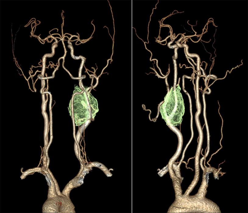
  • Single, smooth pulsatile mass of 3×3 cm present in the right carotid triangle. The skin over the swelling appears stretched. No color changes, discharging sinuses or dilated veins.
  • On palpation, the mass is firm, non-tender with transverse mobility and restricted vertical mobility.
  • The swelling became less prominent on contracting deep cervical fascia and sternocleidomastoid.
  • No other neck swellings palpable.
  • Healed scar present on right side of the neck.
  • Gag reflex present.
  • Rest of the systemic examination was unremarkable.

Questions

  1. What is your provisional diagnosis?


Explanation

Diagnosis is a carotid body tumor.

Above picture is that of a CT Angiogram (arch of the aorta to vertex) showing complete encasement of proximal aspects of right ECA and ICA by the tumor.

Carotid body tumor, also known as a carotid body paraganglioma or chemodectoma are highly vascular tumors arising from the paraganglion cells of the carotid body, located at the site of the carotid bifurcation.

Most common clinical presentation of carotid body tumor is a painless round lateral neck mass, located along the anterior border of upper one-third of the sternocleidomastoid muscle. Involvement of cranial nerves will lead to associated symptoms. Most commonly involved cranial nerves are superior laryngeal, vagus, and hypoglossal nerves.

Digital Subtraction Angiography (DSA) is the gold standard in the diagnosis of carotid body tumors.  The splaying of the carotid vessels (lyre sign) is characteristic of these tumors.

Early and complete surgical resection is often considered as the treatment of choice.  Shamblin classification of carotid body tumors helps in predicting the prognosis and difficulties for surgical resection.

References

Author

Dr. Sanu. P. Moideen, MBBS, MS (ENT), DNB (ENT), FHNOS, is an otolaryngologist (ENT surgeon), head and neck oncosurgeon practising in Muvattupuzha, Kerala, India. After finishing his postgraduate training, he pursued specialist training in paediatric ENT and head and neck oncosurgery from eminent institutions in India and the US.

Leave a Reply

Your email address will not be published. Required fields are marked *