ENT Quiz – Resurgence of a disease of the past

diphtheria

A 6-year-old boy from a rural area presented to pediatric casualty with a history of throat pain, progressive swelling of the neck and moderate grade fever for the past 2 weeks associated with gradually worsening dysphagia for the past 2 days.

Antenatal history was uneventful, born at term by normal vaginal delivery, developmentally appropriate for the age and has no history of any developmental delay. He has not received any immunization to date.

2 days later child developed nasal regurgitation, marked body weakness, mild respiratory distress that progressively became severe, hypotension and ventricular tachycardia.

On examination

  • Awake, toxic looking and febrile.
  • Diffuse swelling of neck involving bilateral submental and jugular nodes.
  • Temperature: 99.5°F, Heart rate: 132/min, Blood pressure: 104/56mmHg, Respiratory rate: 32/min, SpO2: 100% at room air
  • CVS: S1 & S2 Heard, No murmur.
  • RS: Bilateral equal air entry present. Bilateral crepitations and wheeze present.
  • CNS: No focal neurological deficits, No neck stiffness.
  • Abdomen: Soft, nontender. No organomegaly

Local examination

  • Oral cavity: Normal
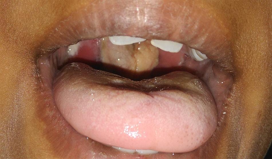
  • Oropharynx: Bilateral Grade IV tonsils with large grayish-white membrane spread over both tonsils extending upto the soft palate was noted. Uvula and soft palate appeared edematous and was pushed anteriorly.

Investigations

  • CRP < 2sec
  • Hemoglobin: 11.1gm%
  • Platelet count: 13,000/cumm
  • Total count: 22,600/cumm, Differentials: N66 / E4 / B0 / L15 / M13
  • ECG: Showing intraventricular block
  • ECHO: Global hypokinesia of left ventricle with an ejection fraction of 45%.

Question

  1. What are the most probable clinical diagnosis and reasons?


Explanation

The clinical diagnosis is faucial diphtheria.

Supporting reasons are non-immunized child, sore throat, enlarged upper deep cervical lymph node, marked weakness, respiratory distress, the extension of the membrane outside the tonsil, low-grade fever with marked tachycardia and leukocytosis.

Diphtheria is caused by a gram-positive bacillus Corynebacterium diptheriae. Diphtheria toxin (exotoxin) is produced by C. diphtheriae only when infected with a bacteriophage that integrates the toxin-encoding genetic elements into the bacteria.

Clinical manifestations

  1. Neck swelling: markedly enlarged (Bull’s Neck) and tender cervical lymphadenopathy is common in the early stages of diphtheria.
  2. Grayish membrane extending outside tonsillar surface: Diphtheria exotoxin causes necrosis of epithelial cells and liberates serous and fibrinous material which forms a grayish-white pseudo-membrane. Thus, the membrane consists of necrotic tissue, bacteria, and rich fibrinous exudate.
  3. As diphtheria is a disease of the mucous membrane, the membrane extends outside the tonsillar surfaces.
  4. Hypotension and ventricular tachycardia were due to diphtheria myocarditis.

More about signs, symptoms, investigations and treatment options of diphtheria can be found here.

Author

Dr. Sanu. P. Moideen, MBBS, MS (ENT), DNB (ENT), FHNOS, is an otolaryngologist (ENT surgeon), head and neck oncosurgeon practising in Muvattupuzha, Kerala, India. After finishing his postgraduate training, he pursued specialist training in paediatric ENT and head and neck oncosurgery from eminent institutions in India and the US.

Leave a Reply

Your email address will not be published. Required fields are marked *