Dirofilaria is a parasitic worm commonly found in dogs and other animals. Human infection, known as dirofilariasis, is relatively uncommon and occurs when the parasite is transmitted through mosquito bites. The majority of human dirofilariasis cases involve pulmonary and subcutaneous lesions.
About 40 types of Dirofilaria species have been identified, and the commonest species which infects humans are Dirofilaria repens and Dirofilaria immitis. In humans, the lung lesions are caused by Dirofilaria immitis while the subcutaneous or subconjunctival lesions are caused mostly by Dirofilaria repens.
However, cases of Dirofilaria affecting deep tissues, such as muscles, are exceptionally rare. This case report describes a unique presentation of dirofilariasis in a 15-year-old girl with a parasitic infection localized in the temporalis muscle.
Case Report:
A 15-year-old girl presented to ENT department with a complaint of a slowly growing swelling in the left temporal region of her head for the past three months. She reported experiencing intermittent dull pain in the same area and noted limited jaw movement, especially when chewing. The patient denied any recent history of travel or exposure to pets. Her medical history was unremarkable, with no history of immunosuppression or chronic illnesses.
On physical examination, a firm, non-tender mass was palpable in the right temporal region, measuring approximately 1.5 cm in diameter. The overlying skin appeared normal, without signs of inflammation or infection. The patient was afebrile, and her vital signs were within normal limits. A thorough neurological examination revealed no abnormalities, except for limited jaw excursion due to discomfort during movement.
Given the patient’s clinical presentation and the location of the swelling, initial differential diagnoses included a benign soft tissue tumor, cyst, or abscess. Blood tests, including complete blood count and inflammatory markers, were within normal ranges. An ultrasound of the temporal region was performed, which revealed a hypoechoic mass with ill-defined margins superficial to temporalis fascia.
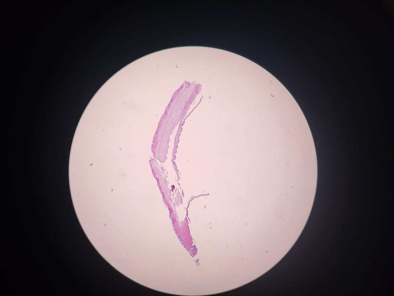
Given the inconclusive findings from the imaging studies, to establish a definitive diagnosis, a surgical excisional biopsy was performed under local anesthesia. Surgical exploration revealed a 2cm distinct firm (indurated) mass adhering to the superficial layer of the temporal muscle, just above the temporalis fascia. The surgical specimen was sent for histopathological examination. Microscopic examination revealed the presence of a coiled, tubular nematode consistent with Dirofilaria species. The nematode displayed fragments Dirofilaria confirming the diagnosis of dirofilariasis.
The literature regarding the medical management of D. repens is limited. Hence after obtaining a consultation with the infectious department, the patient was prescribed a course of oral anti-parasitic medications (Ivermectin 150 to 200 mcg/kg as a single dose) to eliminate any remaining parasites and prevent further complications.
Because of the patient’s discomfort and limited jaw movement, analgesics and anti-inflammatory medications were also prescribed to manage pain and reduce inflammation in the affected temporalis muscle. Non-steroidal anti-inflammatory drugs (NSAIDs) ibuprofen was utilized to alleviate symptoms and improve the patient’s quality of life during the recovery process.
Additional investigations like chest x-ray excluded further manifestations of the parasite. Apart from the routine post op follow up, the patient was reviewed six months after the surgery, and she remains symptom free.