Microtia is a congenital external ear deformity characterized by hypoplasia (incomplete development) of the pinna, ranging from the measurably small external ear with a minimal structural abnormality, to ear with major structural alteration or to a total absence of ear (anotia).
The reported prevalence varies from 0.83 to 17.4 per 10,000 births worldwide. Higher prevalence rates are observed in Hispanics and Asians.
The condition leaves the affected children with a hooked piece of cartilage and skin on one or both sides of their head, along with a host of hearing issues. This can seriously influence the psychological and physiological well-being of affected children.
Treatment options for microtia
Current cosmetic procedures of treating microtia mainly include wearing of auricular prosthesis, implantation of non-absorbable auricular frame materials (such as silastic or high-density polyethylene/ Medpor®), or an autologous rib cartilage framework. Non-absorbable frames generate an excellent ear shape without donor site morbidity, but they lack bioactivity and can lead to extrusion and infections.
Autologous rib cartilage transplantation is currently considered as the gold stand treatment for microtia. But harvesting rib cartilage is associated with donor site injury, and replicating the complex 3D ear structure is hard to achieve.
3D-printed ears and invitro regeneration
In a first-of-its-kind study marking a new milestone in the field of tissue engineering, scientists from Shanghai province China have grown new ears for five children born with microtia, by taking a scan of the child’s unaffected ear, reversing the dimensions and 3D-printing a biodegradable mold punctuated with tiny holes.
In a paper published in the journal EBioMedicine, the team outlines how they developed these 3D-printed ears.
The scientists collected cartilage cells (chondrocytes) from patients’ affected ears to grow new cartilage in vitro. They then 3D-printed mirrored copies of each patient’s healthy ear to make biodegradable molds full of tiny holes. The harvested cartilage cells were then grown inside the mold, ensuring that the new ears would be the right shape for each individual. Over 12 weeks, the cells started to grow into the shape of the mold, replacing bits of it that had already disintegrated. This mold was then grafted onto the children to reconstruct their ear.
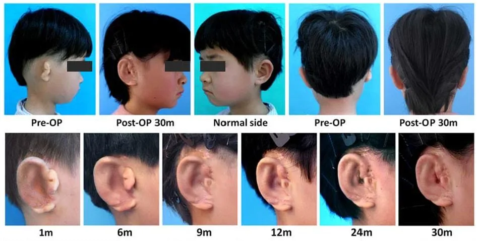
The surgery was done in 5 unilateral microtia children aging from 6 to 10 years. The first surgery was done on a 6-year-old female child and was followed up for 2.5 years.
“The engineered ear graft for each of the total five patients showed excellent cartilage formation in vitro with detailed ear structure, indicating that the procedures for engineering the ear graft were feasible and repeatable. We were able to successfully design, fabricate, and regenerate patient-specific external ears,” wrote the researchers.
This experiment of the 3D-printed ears wasn’t without any negatives though. Two of the patients’ new ears showed slight distortion after surgery. There were many limitations to the research also: growing the cells in culture remains a risky strategy since cells can divide in unpredictable ways, while using the children’s own chondrocytes itself presents problems, since their ears were abnormal and potentially diseased -Tessa Hadlock from the Massachusetts Eye and Ear Infirmary, who wasn’t involved in the study, told CNN.
What’s in the horizon?
The concept of regenerating human pinna in the lab is not a new one. The original concept was first published in 1997, where researchers grew tissue-engineered cartilage in the shape of a human ear using chondrocytes and then implanted them on the backs of mice. From there, it took another long 20 years to have the first successful trial in human beings.
There is another parallel research happening by Professor Bruno Peault, a regenerative medicine expert based at the University of Edinburgh in the U.K. as well as at UCLA. He and his colleagues are working on a similar project using 3D-printed molds to grow new ears from purified stem cells of the patient’s that can be guided into becoming cartilage cells. Peault’s team is still collecting the research needed to win approval for human trials of their method; it might be another one to two years before that might become a reality.
The tissue-engineered auricle is a promising alternative to current ear reconstructive options, but its clinical translation is yet to be accomplished. The method followed by the Chinese scientists is very complicated, expensive, and would be hard to do on a large scale, so it’s unlikely to become widespread. More research is needed before the approach described in the new study could be widely used among microtia patients in a clinical setting.
References
- Zhou G, Jiang H, Yin Z, Liu Y, Zhang Q, Zhang C, Pan B, Zhou J, Zhou X, Sun H, Li D. In Vitro Regeneration of Patient-specific Ear-shaped Cartilage and Its First Clinical Application for Auricular Reconstruction. EBioMedicine. 2018 Jan 13.